图书简介
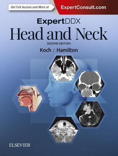
BMA MEDICAL BOOK AWARDS: Now fully revised and up-to-date, Expert DDx: Head and Neck, 2nd edition, quickly guides you to the most likely differential diagnoses based on key imaging findings and clinical information. Expert radiologists Bernadette L. Koch, MD and Bronwyn E. Hamilton, MD present more than 160 cases across a broad spectrum of head and neck diseases, classified by specific anatomic locations, generic imaging findings, modality-specific findings, and clinically based indications. Readers will find authoritative, superbly illustrated guidance for defining and reporting useful, actionable differential diagnoses that lead to definitive findings in every area of the head and neck. Presents at least eight clear, sharp, succinctly annotated images for each diagnosis (more than 2,500 annotated images in all); a list of diagnostic possibilities sorted as common, less common, and rare but important; and brief, bulleted text offering helpful diagnostic clues Shows both typical and variant manifestations of each possible diagnosis Includes new cases, expanded differential considerations, new references, and updated imaging throughout Covers hot topics such as the evolving role of imaging with respect to many head and neck conditions, new ACR white paper recommendations on incidental thyroid nodule work-up, an expanding number of recognized genetic and syndromic diseases, updated information about IgG-4 related disease imaging manifestations in the head
Koch and Hamilton, Expert DDx Head and Neck Suprahyoid & Infrahyoid Anatomically Based Differentials Parapharyngeal Space Lesion Pharyngeal Mucosal Space Lesion, Nasopharynx Pharyngeal Mucosal Space Lesion, Oropharynx Masticator Space Lesion Buccal Space Lesion Parotid Space Mass Carotid Space Lesion Carotid Artery Lesion Perivertebral Space Lesion Brachial Plexus Lesion Visceral Space Lesion Cervical Tracheal Lesion Tracheoesophageal Groove Lesion Posterior Cervical Space Lesion Cervicothoracic Junction Lesion TMJ Mass Lesions Calcified TMJ Lesions TMJ Cysts Generic Imaging Patterns Diffuse Parotid Disease Multiple Parotid Masses Focal Retropharyngeal Space Mass Diffuse Retropharyngeal Space Disease Diffuse Thyroid Enlargement Focal Thyroid Mass Invasive Thyroid Mass Clinically Based Differentials Cheek Mass Trismus Oral Cavity, Mandible & Maxilla Anatomically Based Differentials Oral Mucosal Space/Surface Lesion Sublingual Space Lesion Submandibular Space Lesion Submandibular Gland Lesion Root of Tongue Lesion Hard Palate Lesion Maxillary Bone Lesion Generic Imaging Patterns Tooth-Related Mass, Sclerotic Tooth-Related Mass, Cystic Modality-Specific Imaging Findings Low-Density Jaw Lesion, Sharply Marginated (CT) Low-Density Jaw Lesion, Poorly Marginated (CT) Ground-Glass Lesions of Mandible & Maxilla (CT) Hypopharynx & Larynx Anatomically Based Differentials Hypopharyngeal Lesion Laryngeal Lesion Generic Imaging Patterns Epiglottic Enlargement Diffuse Laryngeal Swelling Subglottic Stenosis Clinically Based Differentials Vocal Cord Paralysis (Left) Vocal Cord Paralysis (Right) Stridor in Child Lymph Nodes Generic Imaging Patterns Enlarged Lymph Nodes in Neck in Adult Avidly Enhancing Lymph Nodes Enlarged Lymph Nodes in Neck of Child Transspatial, Multispatial, or Multilocation in Head and Neck Generic Imaging Patterns Air-Containing Lesions in Neck Solid Neck Mass in Infant Solid Neck Mass in Child Cystic Neck Mass in Child Cystic-Appearing Neck Masses in Adult Transspatial Mass in Child Transspatial Neck Mass Modality-Specific Imaging Findings Hyperdense Neck Lesion (CT) Low-Density Neck Lesion (CT) Hypervascular Neck Lesion (CT/MR) Clinically Based Differentials Angle of Mandible Mass Supraclavicular Mass Sinus and Nose Anatomically Based Differentials Sinonasal Anatomic Variants Generic Imaging Patterns Nasal Septal Perforation Congenital Midline Nasal Lesion Fibroosseous & Cartilaginous Lesions Inflammatory Patterns of Sinusitis Multiple Sinonasal Lesions Expansile Sinonasal Lesion Nasal Lesion With Bone Destruction Nasal Lesion Without Bone Destruction Sinus Lesion Without Bone Destruction Sinus Lesion With Bone Destruction Facial Bone Lesion Modality-Specific Imaging Findings Hyperdense Disease in Sinus Lumen (CT) Calcified Sinonasal Lesion (CT) T2 Hypointense Sinus Lesion (MR) Clinically Based Differentials Nasal Obstruction in Newborn Anosmia-Hyposmia Epistaxis Traumatic Lesions of Face Orbit Anatomically Based Differentials Preseptal Lesion Ocular Lesion, Adult Ocular Lesion, Child Optic Nerve-Sheath Lesion Intraconal Mass Extraconal Mass Lacrimal Gland Lesion Orbital Wall Lesion Generic Imaging Patterns Microphthalmos Macrophthalmos Optic Nerve Sheath Tram-Track Sign Extraocular Muscle Enlargement Large Superior Ophthalmic Vein(s) Ill-Defined Orbital Mass Cystic Orbital Lesions Vascular Lesions of Orbit Accidental Foreign Bodies in Orbit Surgical Devices & Treatment Effects in Orbit Modality-Specific Imaging Findings Intraocular Calcifications (CT) Clinically Based Differentials Leukocoria Painless Proptosis in Adult Painful Proptosis in Adult Rapidly Developing Proptosis in Child Infectious & Inflammatory Orbital Lesions Extraocular Orbital Mass in Child Temporal Bone Anatomically Based Differentials External Auditory Canal Lesion Middle Ear Lesion, Child Middle Ear Lesion, Adult Inner Ear Lesion, Adult Petrous Apex Lesion Inner Ear Lesion, Child Facial Nerve Lesion, Temporal Bone Generic Imaging Patterns Enhancing Middle Ear Lesions Expansile-Destructive Petrous Apex Lesion Bony Lesions of Temporal Bone Clinically Based Differentials Conductive Hearing Loss Peripheral Facial Nerve Paralysis Vascular Retrotympanic Mass Pulsatile Tinnitus Traumatic Lesions of Temporal Bone Secondary (Referred) Otalgia Skull Base Anatomically Based Differentials Normal Skull Base Venous Variants Skull Base Foraminal or Canal Variants Congenital Anomalies of Skull Base Intrinsic Skull Base Lesion Anterior Skull Base Lesion Cribriform Plate Lesion Central Skull Base Lesion Sellar/Parasellar Mass With Skull Base Invasion Unilateral Cavernous Sinus Mass Bilateral Cavernous Sinus Mass Meckel Cave Lesion Posterior Skull Base Lesion Clival Lesion Jugular Foramen Lesion Dural Sinus Lesion, General Foramen Magnum Mass CPA-IAC & Posterior Fossa Anatomically Based Differentials Small Internal Auditory Canal Large Internal Auditory Canal CPA Mass, Adult Prepontine Cistern Mass Cisterna Magna Mass Posterior Fossa Neoplasm, Adult Posterior Fossa Neoplasm, Pediatric Generic Imaging Patterns Cystic CPA Mass Clinically Based Differentials Hemifacial Spasm Sensorineural Hearing Loss in Adult Sensorineural Hearing Loss in Child Cranial Nerve & Brainstem Anatomically Based Differentials Midbrain Lesion Pontine Lesion Medulla Lesion Clinically Based Differentials Monocular Vision Loss Bitemporal Heteronymous Hemianopsia Homonymous Hemianopsia Oculomotor, Trochlear, or Abducens Neuropathy Trigeminal Neuropathy Trigeminal Neuralgia Complex Cranial Nerve IX-XII Neuropathy Hypoglossal Neuropathy Horner Syndrome Generic Imaging Patterns Enhancing Cranial Nerve(s)
Trade Policy 买家须知
- 关于产品:
- ● 正版保障:本网站隶属于中国国际图书贸易集团公司,确保所有图书都是100%正版。
- ● 环保纸张:进口图书大多使用的都是环保轻型张,颜色偏黄,重量比较轻。
- ● 毛边版:即书翻页的地方,故意做成了参差不齐的样子,一般为精装版,更具收藏价值。
关于退换货:- 由于预订产品的特殊性,采购订单正式发订后,买方不得无故取消全部或部分产品的订购。
- 由于进口图书的特殊性,发生以下情况的,请直接拒收货物,由快递返回:
- ● 外包装破损/发错货/少发货/图书外观破损/图书配件不全(例如:光盘等)
并请在工作日通过电话400-008-1110联系我们。
- 签收后,如发生以下情况,请在签收后的5个工作日内联系客服办理退换货:
- ● 缺页/错页/错印/脱线
关于发货时间:- 一般情况下:
- ●【现货】 下单后48小时内由北京(库房)发出快递。
- ●【预订】【预售】下单后国外发货,到货时间预计5-8周左右,店铺默认中通快递,如需顺丰快递邮费到付。
- ● 需要开具发票的客户,发货时间可能在上述基础上再延后1-2个工作日(紧急发票需求,请联系010-68433105/3213);
- ● 如遇其他特殊原因,对发货时间有影响的,我们会第一时间在网站公告,敬请留意。
关于到货时间:- 由于进口图书入境入库后,都是委托第三方快递发货,所以我们只能保证在规定时间内发出,但无法为您保证确切的到货时间。
- ● 主要城市一般2-4天
- ● 偏远地区一般4-7天
关于接听咨询电话的时间:- 010-68433105/3213正常接听咨询电话的时间为:周一至周五上午8:30~下午5:00,周六、日及法定节假日休息,将无法接听来电,敬请谅解。
- 其它时间您也可以通过邮件联系我们:customer@readgo.cn,工作日会优先处理。
关于快递:- ● 已付款订单:主要由中通、宅急送负责派送,订单进度查询请拨打010-68433105/3213。
本书暂无推荐
本书暂无推荐